Skip to content Skip to sidebar Skip to footer

Foot Ct Scan Position

Ct Scan Patient Instruction 24 Lower Limb Ankle Foot Flexion Extension Youtube

Ct Scan Patient Instruction 24 Lower Limb Ankle Foot Flexion Extension Youtube

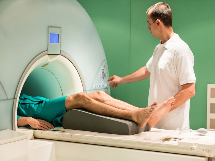
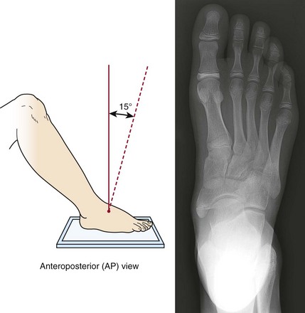
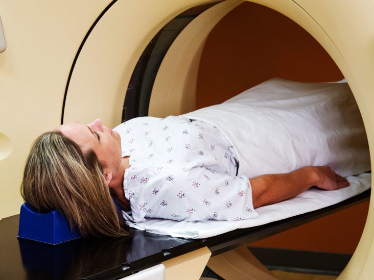
Foot ct scan position. Imaging is performed with the patient in prone. The foot is typically held in a neutral position and sagittal and axial images are taken of the tendon. And even the positioning of your foot and the relief you get when you hold your foot in a different position can tells a lot about whether or not an MRI or CT scan will be helpful for you.

SPECT high resolution CT images left to right. Color sonography can also been used to detect posterior tibial tendinopathy. Although there are methods to simulate the standing position while the patient is lying on a table the best way to evaluate foot and ankle biomechanics is to have the patient stand.

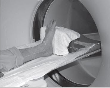
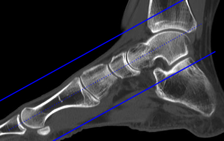
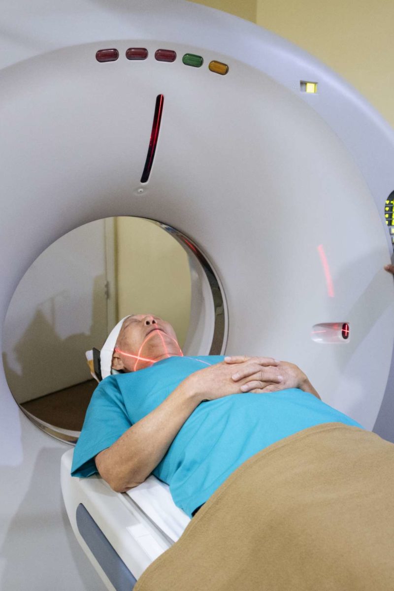
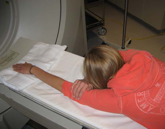
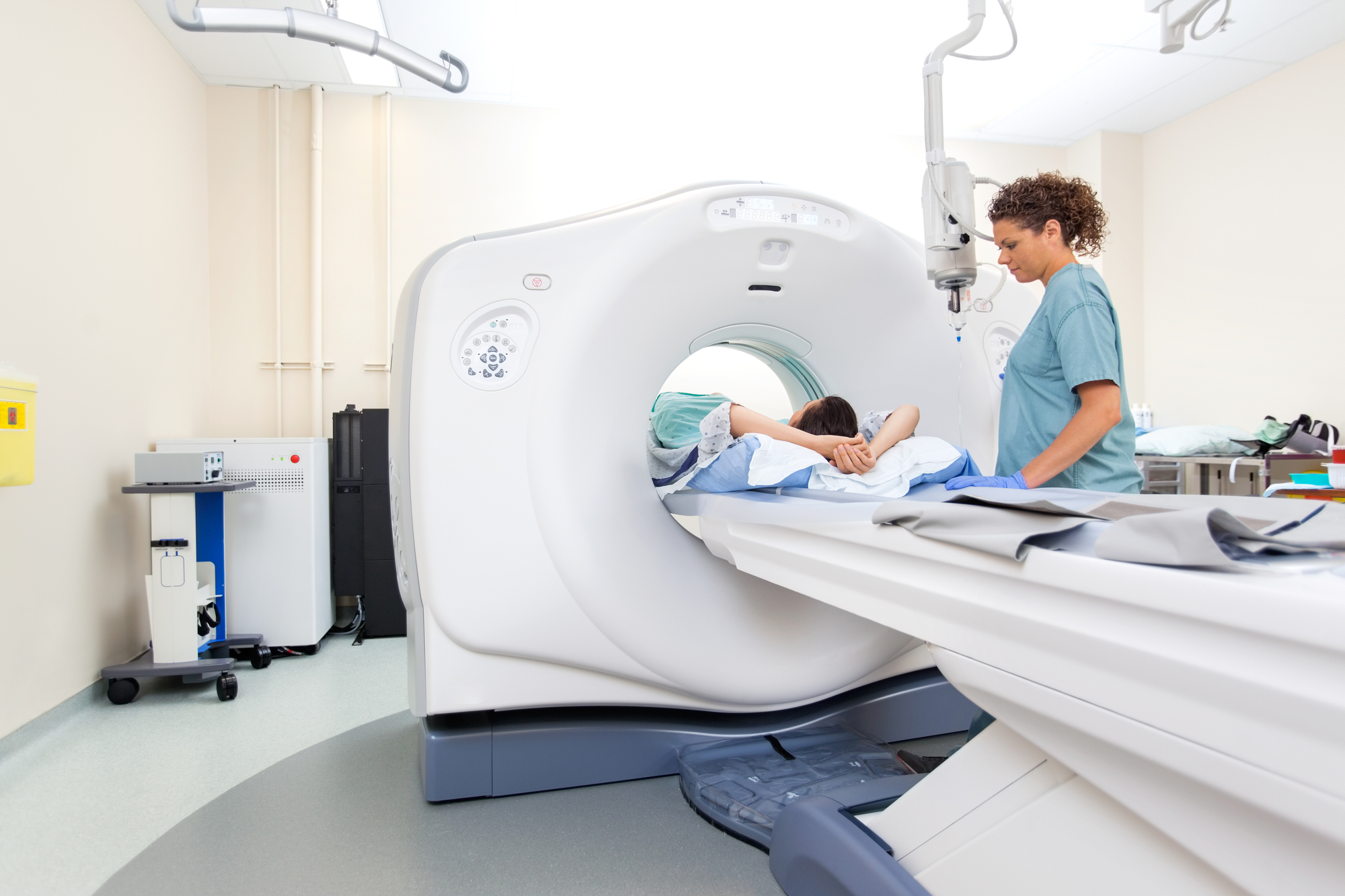
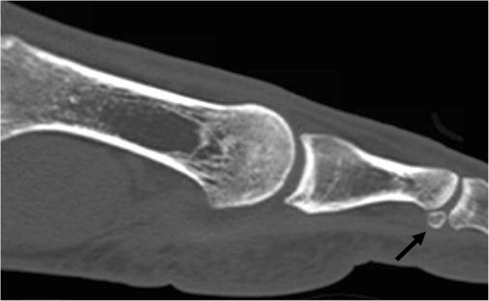
Transverse sagittal and coronal of the left foot of a 21-year-old female with an os tibiale externum in both feet and a history of fixation of the os tibiale externum at the left side with persistent painSPECT-CT shows high focal activity in the os tibiale externum at the left side indicative of nonunion. Relevant Anatomy C a l c a n e u s C u b o i d L a t e r a l C u n e i f o r m M e d i a l C u n e i f o r m I n t r m e d i a t e u n e f o r m. Patient Supine with feet first into scanner Keep knees extended side- by-side.

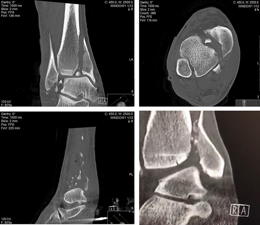
They also show soft tissues such as cartilage ligaments and muscles more clearly than traditional X-rays so they are more useful for diagnosing. The foot is prone to several types of damage and injuries. Make mortise coronal and sagittal MPRs as depicted below using thin data set with sharp kernel.

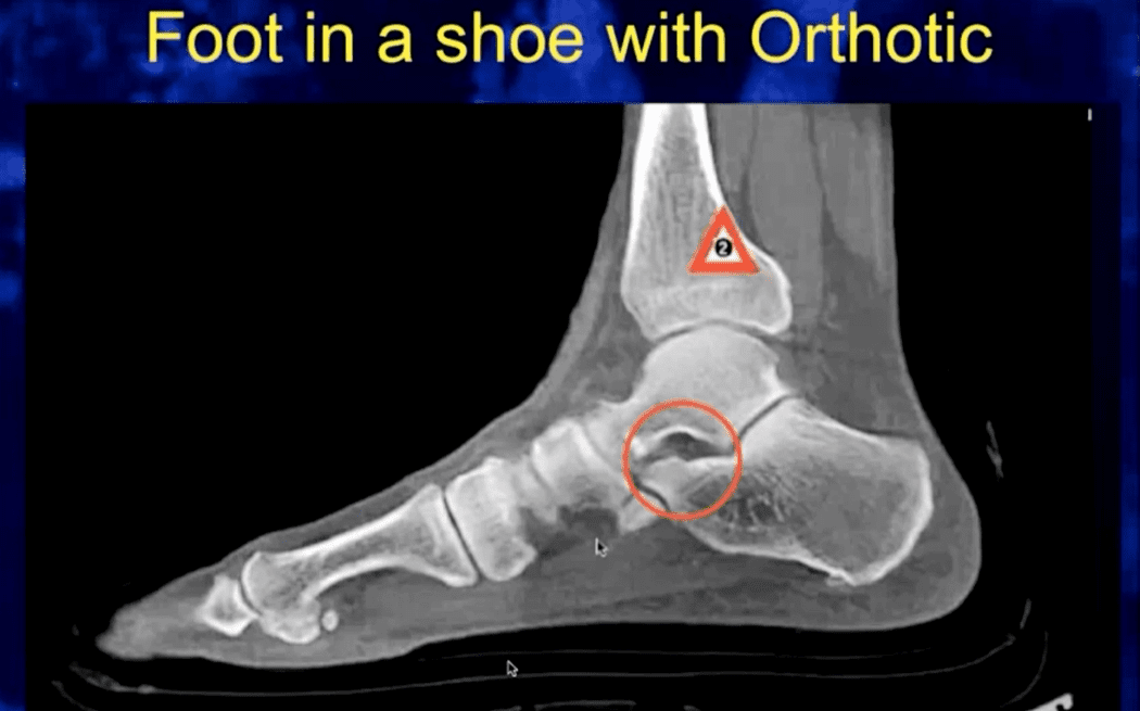
CT scans help doctors distinguish between a simple cyst and a solid tumor and any involvement of the bone. In a traditional CT scan the foot is in a relaxed position. Above tibiafibula joint through hind foot.

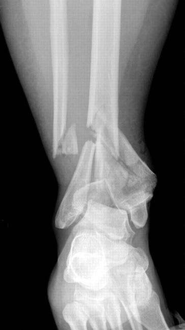
Foot and Ankle CT Scan CT scans may be used to diagnose ankle fractures that dont show up on X-ray. Pain and inflammation of the foot may cause limited movement 5. Although there are methods to simulate the standing position while the patient is lying on a table the best way to evaluate foot and ankle biomechanics is to have the patient stand.

In a traditional CT scan the foot is in a relaxed position. Positioning Patient supine.

Ankle And Foot Ct Imaging Technique Radtechonduty

Ankle And Foot Ct Imaging Technique Radtechonduty

In Office Foot Ct Scan Dr Paul Drucker Podiatrist Ny

In Office Foot Ct Scan Dr Paul Drucker Podiatrist Ny

Ct Scan Of Foot Positioning Ct Scan Machine

Ct Scan Of Foot Positioning Ct Scan Machine

2

2

Ct Scan Foot Positioning Page 1 Line 17qq Com

Ct Scan Foot Positioning Page 1 Line 17qq Com

Msk Ct Protocols

Msk Ct Protocols

1 Position And Orientation Of Lower Extremity Sample In Styrofoam Download Scientific Diagram

1 Position And Orientation Of Lower Extremity Sample In Styrofoam Download Scientific Diagram

Foot And Ankle Ct Scan Omega Pds

Foot And Ankle Ct Scan Omega Pds

2

2

Pedcat Ct Scans Foot Ankle Deformities Pittsburgh Pa

Pedcat Ct Scans Foot Ankle Deformities Pittsburgh Pa

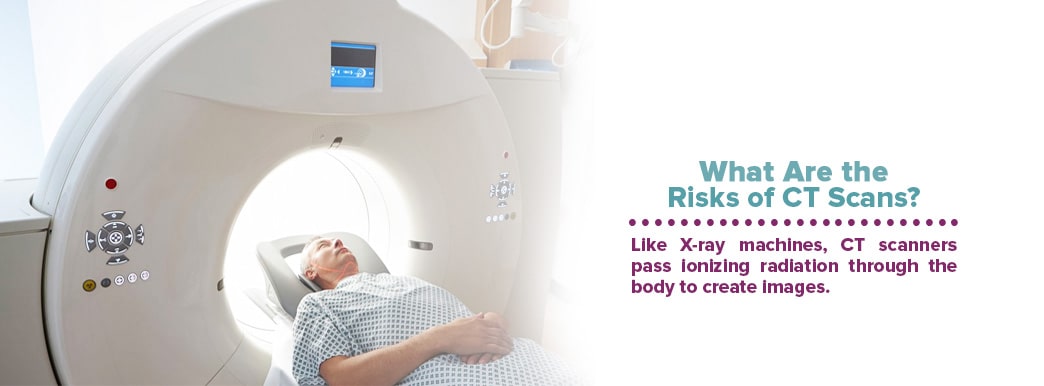
Ct Scan Or Cat Scan How Does It Work

Ct Scan Or Cat Scan How Does It Work

Webinar Recap Using Weight Bearing Ct To Guide Clinical Decisions Curvebeam

Webinar Recap Using Weight Bearing Ct To Guide Clinical Decisions Curvebeam

Computed Tomography Ct Scanning Of An Ankle In Full Plantar Flexion Download Scientific Diagram

Computed Tomography Ct Scanning Of An Ankle In Full Plantar Flexion Download Scientific Diagram

Fast Integrated Workflow

Fast Integrated Workflow

Knee Mri Scan Purpose Procedure And Risks

Knee Mri Scan Purpose Procedure And Risks

Pedcat 3d Weight Bearing Ct University Foot And Ankle Institute

Pedcat 3d Weight Bearing Ct University Foot And Ankle Institute

Ct Scan Elbow Joint Positioning Ct Scan Machine

Ct Scan Elbow Joint Positioning Ct Scan Machine

Msk Ct Protocols

Msk Ct Protocols

Lateral Sesamoid Position Relative To The Second Metatarsal In Feet With And Without Hallux Valgus A Prospective Study The Journal Of Foot And Ankle Surgery

Lateral Sesamoid Position Relative To The Second Metatarsal In Feet With And Without Hallux Valgus A Prospective Study The Journal Of Foot And Ankle Surgery

Mri Scans Physiopedia

Mri Scans Physiopedia

New Imaging Technique Standing Ct Scanning Available At The Oving Clinic Chichester Foot And Ankle

New Imaging Technique Standing Ct Scanning Available At The Oving Clinic Chichester Foot And Ankle

Weight Bearing Cone Beam Ct Scans In The Foot And Ankle Efort Open Reviews

Weight Bearing Cone Beam Ct Scans In The Foot And Ankle Efort Open Reviews

X Rays Ct Scans And Mris Orthoinfo Aaos

X Rays Ct Scans And Mris Orthoinfo Aaos

1

1

Contrast Dye Used For X Rays And Cat Scans

Contrast Dye Used For X Rays And Cat Scans

Ct Computed Tomography Scan What To Expect Youtube

Ct Computed Tomography Scan What To Expect Youtube

Ct Scan Wikipedia

Ct Scan Wikipedia

Diagnostic Imaging Of The Foot And Ankle For Physical Therapists Physiopedia

Diagnostic Imaging Of The Foot And Ankle For Physical Therapists Physiopedia

2

2

Imaging Of The Foot And Ankle Musculoskeletal Key

Imaging Of The Foot And Ankle Musculoskeletal Key

Computed Tomography Ct Angiography Angiogram

Computed Tomography Ct Angiography Angiogram

Foot Mri Protocols And Planning Indications For Mri Foot Scan

Foot Mri Protocols And Planning Indications For Mri Foot Scan

What To Expect During An Extremity Mri Rai Health Awareness Blog

What To Expect During An Extremity Mri Rai Health Awareness Blog

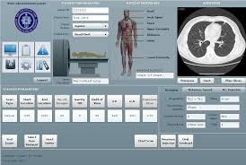
What Is Localizer On Ct Scan Radtechonduty

What Is Localizer On Ct Scan Radtechonduty

Intra Operative 3 Dimensional Imaging O Arm In Foot And Ankle Trauma Surgery Report Of 2 Cases And Review Of The Literature

Intra Operative 3 Dimensional Imaging O Arm In Foot And Ankle Trauma Surgery Report Of 2 Cases And Review Of The Literature

Diagnostic Imaging For Heel Pain X Ray Mri Ultrasound Ct Scan

Diagnostic Imaging For Heel Pain X Ray Mri Ultrasound Ct Scan

Anatomical Variation In The Ankle And Foot From Incidental Finding To Inductor Of Pathology Part Ii Midfooot And Forefoot Insights Into Imaging Full Text

Anatomical Variation In The Ankle And Foot From Incidental Finding To Inductor Of Pathology Part Ii Midfooot And Forefoot Insights Into Imaging Full Text

Msk Ct Protocols

Msk Ct Protocols

Ct And Mr Imaging Of The Postoperative Ankle And Foot Radiographics

Ct And Mr Imaging Of The Postoperative Ankle And Foot Radiographics

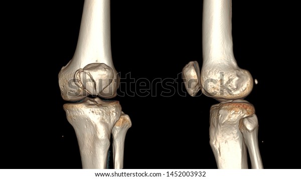
Ct Scan 3d Right Knee Ap Stock Illustration 1452003932

Ct Scan 3d Right Knee Ap Stock Illustration 1452003932

Epos Trade

Epos Trade

X Rays Ct Scans And Mris Orthoinfo Aaos

X Rays Ct Scans And Mris Orthoinfo Aaos

Diagnosing Muscle And Bone Disorders With Ct Scans

Diagnosing Muscle And Bone Disorders With Ct Scans

Weight Bearing Cone Beam Ct Scans In The Foot And Ankle Efort Open Reviews

Weight Bearing Cone Beam Ct Scans In The Foot And Ankle Efort Open Reviews

What Is A Ct Urogram Procedure Uses Risks And Results

What Is A Ct Urogram Procedure Uses Risks And Results

2

2

Lumbar Mri Scan Purpose Procedure And Risks

Lumbar Mri Scan Purpose Procedure And Risks

A Closer Look At Imaging Options For Complicated Heel Pain Podiatry Today

A Closer Look At Imaging Options For Complicated Heel Pain Podiatry Today

1

1

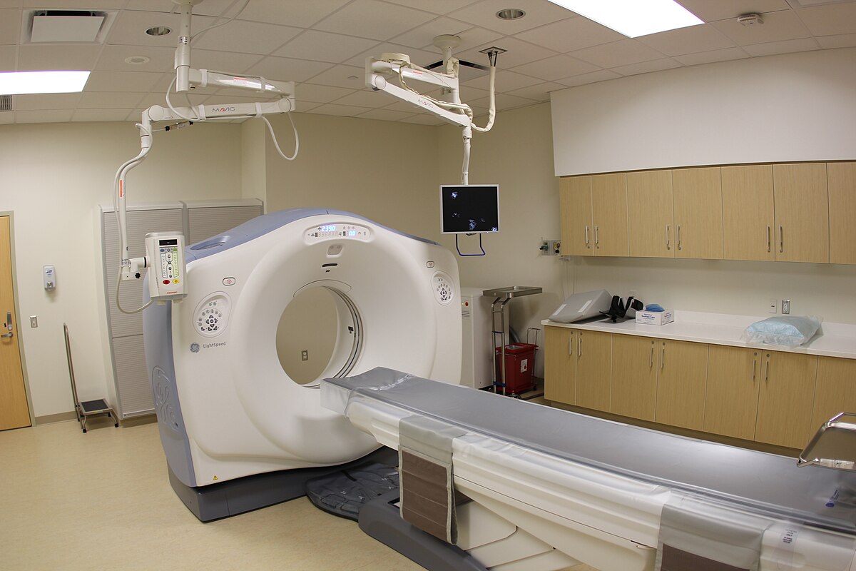
Medical professionals use advanced imaging procedures to diagnose medical problems in the foo t.

Color sonography can also been used to detect posterior tibial tendinopathy. Positioning Patient supine. In a traditional CT scan the foot is in a relaxed position. Microsoft PowerPoint - MSK CT PROTOCOL2 Author. A computed tomography scan of the foot and ankle may help doctors examine any injury or fractures 6. Although there are methods to simulate the standing position while the patient is lying on a table the best way to evaluate foot and ankle biomechanics is to have the patient stand. The foot is typically held in a neutral position and sagittal and axial images are taken of the tendon. And even the positioning of your foot and the relief you get when you hold your foot in a different position can tells a lot about whether or not an MRI or CT scan will be helpful for you. A CT scan of the foot allows physicians to examine bones soft tissues and joints to determine whether there are any damages fractures or any further abnormalities.


7212015 100450 AM. Scan through entire foot. The foot is typically held in a neutral position and sagittal and axial images are taken of the tendon. Imaging is performed with the patient in prone. And even the positioning of your foot and the relief you get when you hold your foot in a different position can tells a lot about whether or not an MRI or CT scan will be helpful for you. Computed tomography offers some advantages over other x-ray techniques in diagnosing disease particularly because it clearly shows the shape and exact location of soft tissues and bones in any slice of the foot and ankle. Although there are methods to simulate the standing position while the patient is lying on a table the best way to evaluate foot and ankle biomechanics is to have the patient stand.

Post a Comment for "Foot Ct Scan Position"