Interstitial Lung Disease Hrct
Interstitial lung disease hrct. Interstitial lung diseases- HRCT 1. Interstitial lung disease ILD is observed in 565 of polymyositis PM and dermatomyositis DM cases 1 2 and is a significant prognostic factor. Interstitial lung disease guideline.
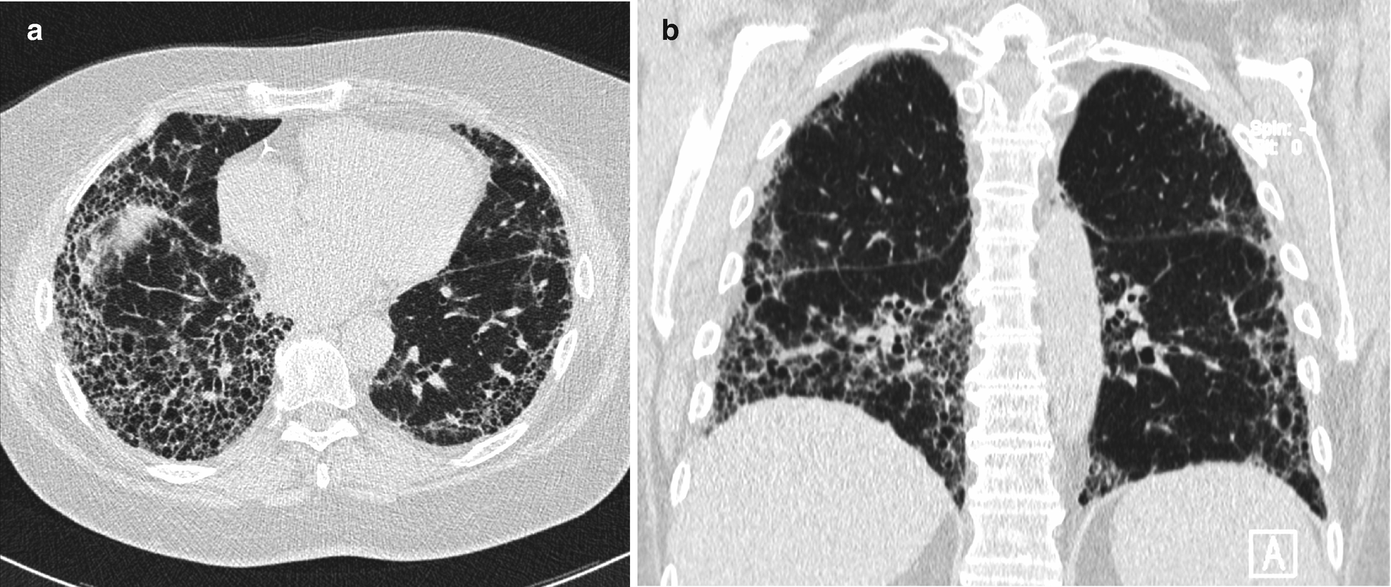
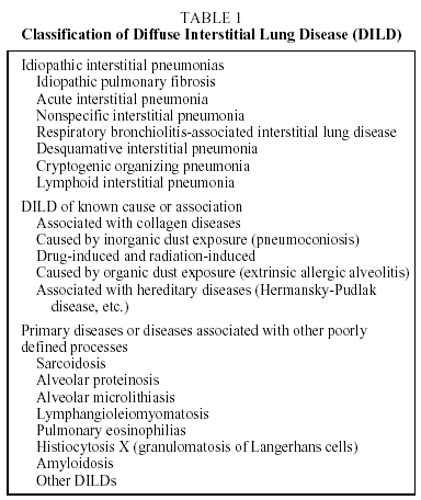
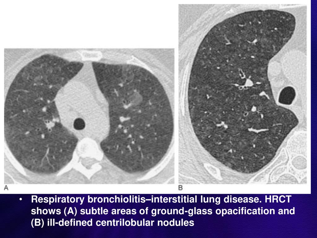
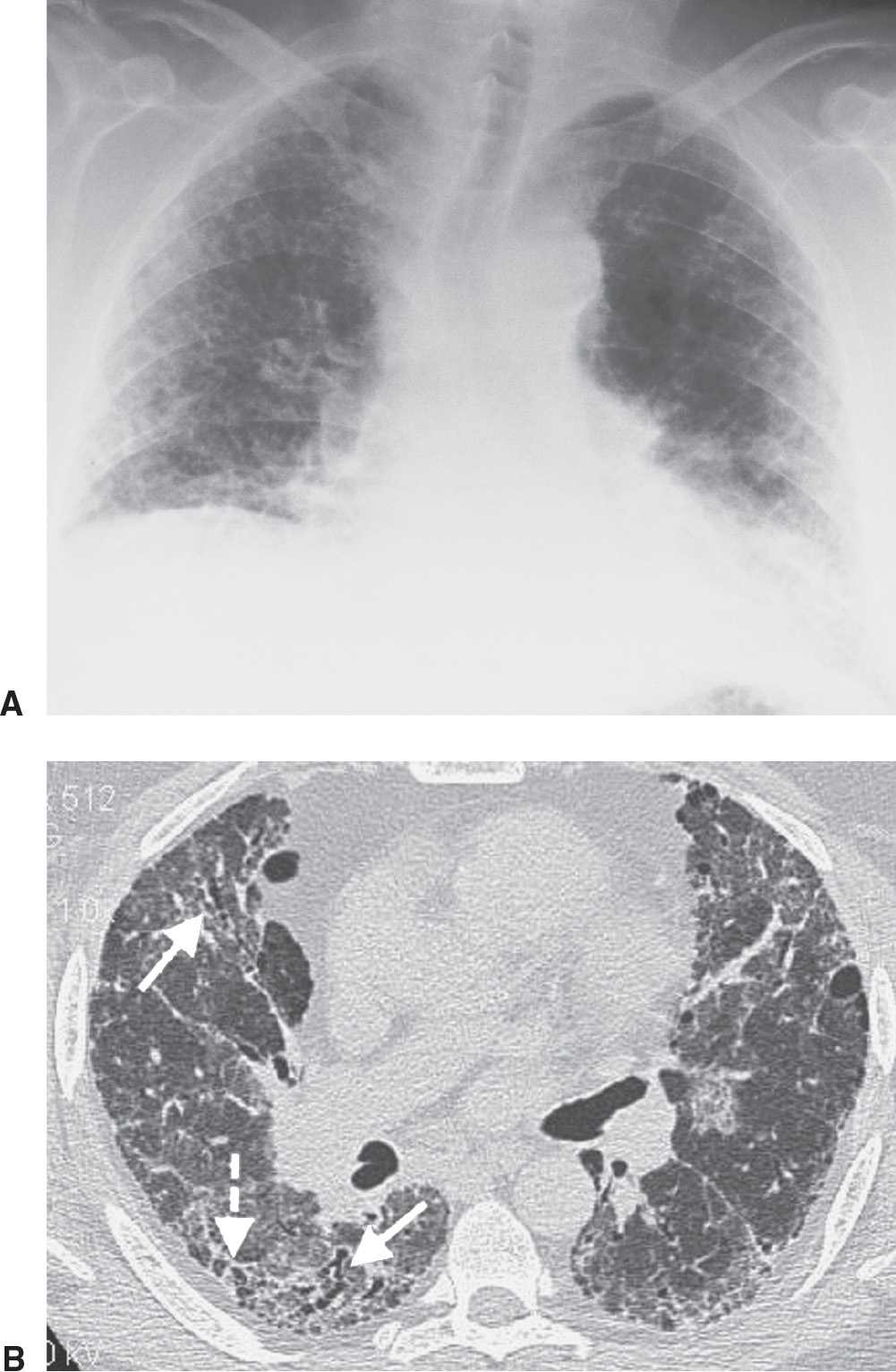
Although the position of HRCT as the dominant imaging technique for interstitial lung disease has remained unchallenged since its introduction in the late 1980s the roles assumed by HRCT have undergone a. The 2018 revised diagnostic HRCT criteria for usual interstitial pneumonia UIP pattern published by the American Thoracic Society the European Respiratory Society the Japanese Respiratory Society and the Latin American Thoracic Association has converged to a similar categorization of the HRCT findings into four groups. Interstitial lung disease is a broad term for a number of diseases that lead to inflammation or scarring of the lungs leading to fibrosis.
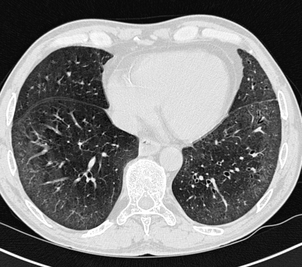
1 PMDM-associated ILD PMDM-ILD can be divided into acute and chronic types. HRCT in the management of diffuse and interstitial lung diseases establish the correct diagnosis or differential diagnosis. HRCT has transformed the diagnostic landscape by providing detailed cross-sectional imaging of the lungs which permit ready identification of a variety of different interstitial lung diseases.
Interstitial Lung Diseases Related to Connective Tissue Disorders Pathology 1. Lung disease a definitive diagnosis being established by specific combinations of clinical radiological and pathological findings. RETICULAR - caused by thickened interlobular or intralobular septa.
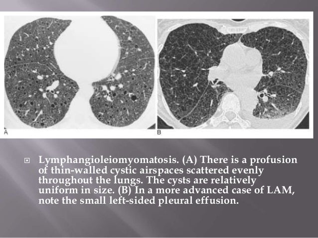
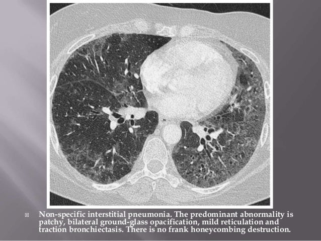
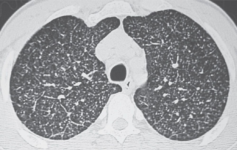
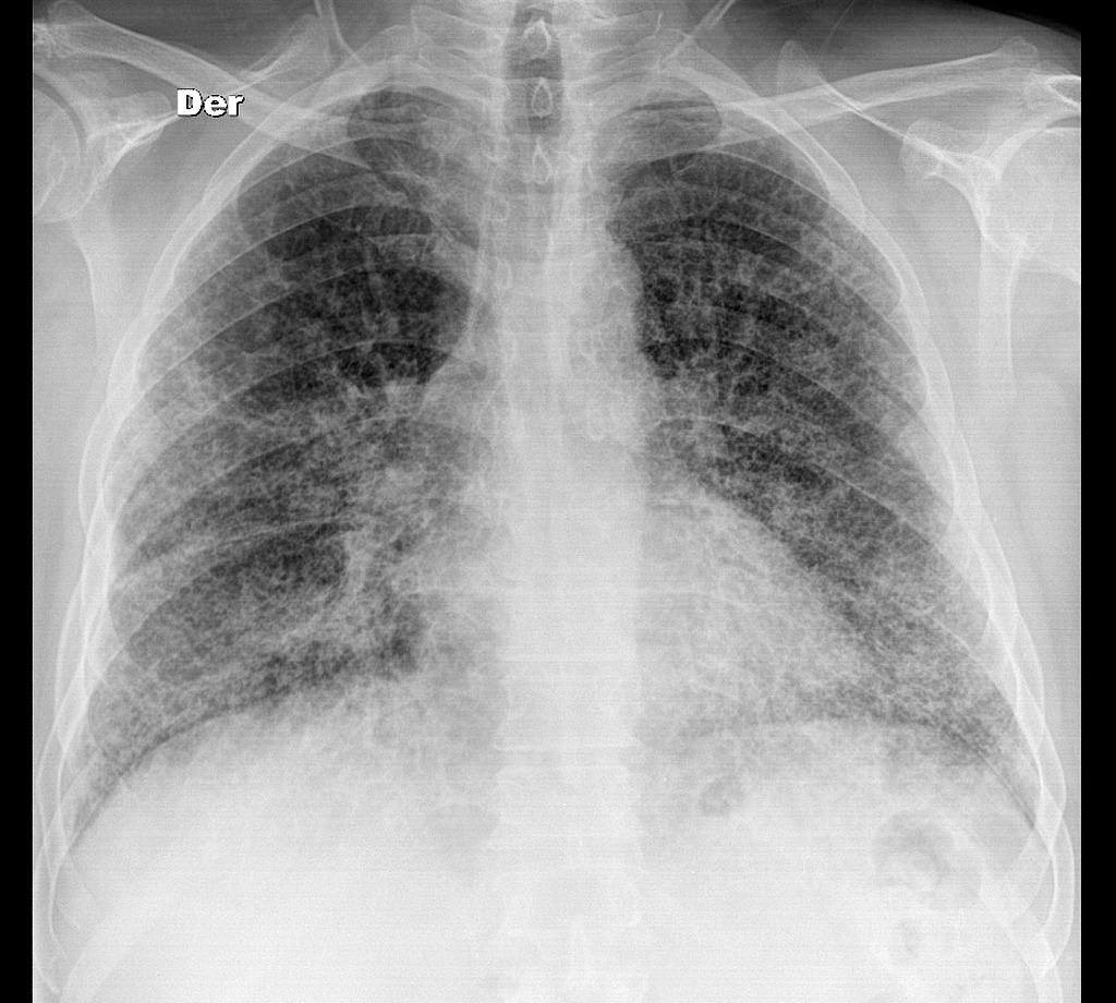
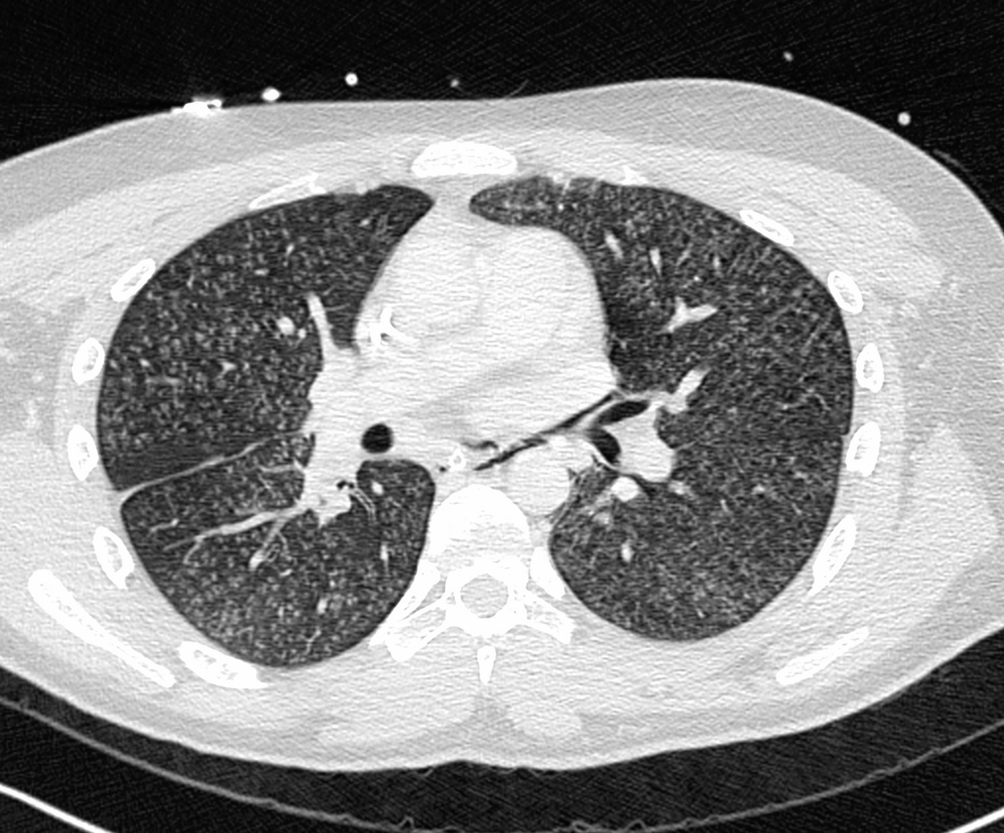
Low attenuation linear opacities nodular and high attenuation. The second part is organized according to the four dominant types of HRCT pattern encountered in interstitial lung disease. Approximately 60 to 70 of patients with sarcoidosis have characteristic radiologic findings.
The differential diagnosis of HRCTs is based on the analysis of the predominant CT pattern the ancillary CT findings and the distribution of the findings. The first section of this brief overview of the value of CT in the diagnostic workup of interstitial lung diseases ILD discusses the technical aspects and the term high-resolution CT HRCT. The concept of separate but connected components making up the lung interstitium propounded by Weibel 17 is important to the understanding of HRCT findings in interstitial lung disease the equivalent terms applicable to HRCT are given in parenthesis.
The HRCT appearance of pulmonary sarcoidosis varies greatly and is known to mimic many other diffuse infiltrative lung diseases. The British Thoracic Society in collaboration with the Thoracic Society of Australia and New Zealand and the Irish Thoracic Society A U Wells1 N Hirani2 on behalf of the British Thoracic Society Interstitial Lung Disease Guideline Group a subgroup of the British Thoracic Society Standards of Care.
However 15 of patients taking docetaxel may develop severe pneumotoxicity.
We report seven breast cancer patients who developed docetaxelinduced diffuse parenchymal lung disease DPLD of an organizing pneumonia pattern on highresolution computed tomography HRCT. Image reconstruction with high spatial resolution. The HRCT appearance of pulmonary sarcoidosis varies greatly and is known to mimic many other diffuse infiltrative lung diseases. The British Thoracic Society in collaboration with the Thoracic Society of Australia and New Zealand and the Irish Thoracic Society A U Wells1 N Hirani2 on behalf of the British Thoracic Society Interstitial Lung Disease Guideline Group a subgroup of the British Thoracic Society Standards of Care. According to current international guidelines HRCT plays a key role in establishing a diagnosis of usual interstitial pneumonia UIP. Radiographic assessment of interstitial lung disease ILD progression and proteins from bronchoalveolar lavage may independently provide better insight. The next section explains when. In 25 to 30 of cases the radiologic findings are atypical. Low attenuation linear opacities nodular and high attenuation.
Knowledge of the lung anatomy is essential for understanding HRCT. It is the smallest lung unit that is surrounded by connective tissue septa. According to current international guidelines HRCT plays a key role in establishing a diagnosis of usual interstitial pneumonia UIP. The second part is organized according to the four dominant types of HRCT pattern encountered in interstitial lung disease. The first section of this brief overview of the value of CT in the diagnostic workup of interstitial lung diseases ILD discusses the technical aspects and the term high-resolution CT HRCT. 3 The acute type of PMDM-ILD is often rapidly progressive and refractory to treatment resulting in fatal outcome. The next section explains when.















Post a Comment for "Interstitial Lung Disease Hrct"